
Skull Base Tumors: Why Specialized Surgical Expertise Makes All the Difference
October 15th, 2026
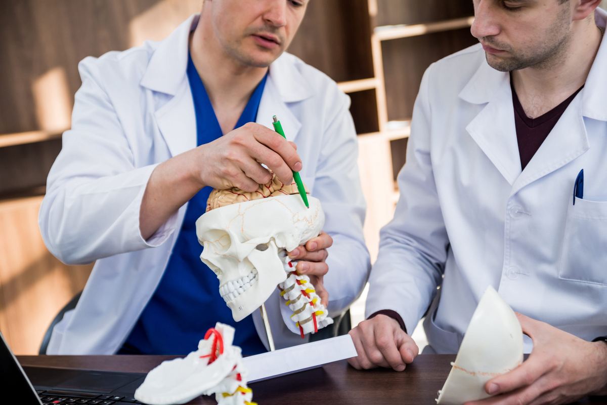
A skull base tumor is a growth in one of the most tightly packed and unforgiving spaces in the human body. The skull base is the bone at the bottom of the skull: the floor that separates the brain from the eyes, nose, sinuses, mouth, and throat. Tumors here sit within millimeters of the optic nerves, the carotid arteries, the brainstem, and the twelve cranial nerves that control vision, facial movement, hearing, swallowing, and speech.
Removing a tumor from that space without damaging the structures around it is one of the most demanding operations in modern surgery. It is also one where the difference between an experienced skull base team and a general surgical approach shows up clearly: in tumor removal, in complications, and in the functions a patient keeps afterward.
This guide explains what skull base surgery involves, why the anatomy makes it so difficult, and what to look for in the team that treats you.
What Is the Skull Base?
Picture the bottom of the skull as an uneven floor with three levels, each lower than the last, sloping from front to back. The front portion sits above the eyes and nasal cavity. The middle portion holds the pituitary gland and the openings for the optic nerves and the carotid arteries. The rear portion contains the brainstem, the spinal cord, and the openings for the nerves that control the face, throat, and tongue.
Almost nothing about this area is spacious. Nerves, arteries, veins, and the brain itself pass through the same narrow corridors, and a tumor the size of a grape can press on several critical structures at once. That compression is what produces symptoms (vision changes, facial numbness, hearing loss, headaches, or hormonal problems) long before the tumor itself becomes dangerous.
Types of Skull Base Tumors
Skull base tumors are not a single disease. They range from slow-growing benign tumors to aggressive cancers, and the type determines almost everything about treatment.
- Meningioma: Arises from the membranes covering the brain. Usually benign and slow-growing, but can compress nerves and vessels as it enlarges.
- Pituitary adenoma: Grows in the pituitary gland at the center of the skull base. Often managed with medication or surgery through the nose.
- Schwannoma: A benign tumor of the nerve sheath, frequently affecting the nerve that controls hearing and balance.
- Chordoma and chondrosarcoma: Rare tumors of the bone and cartilage at the skull base. Locally aggressive and requiring specialized expertise.
- Sinonasal and nasopharyngeal cancers: Malignancies that begin in the nasal cavity, sinuses, or upper throat and extend upward into the skull base.
- Esthesioneuroblastoma: A rare cancer arising high in the nasal cavity, close to the brain.
- Metastatic tumors: Cancer that has spread to the skull base from elsewhere in the body.
Benign does not always mean harmless. A benign tumor pressing on the optic nerve can still take a patient's vision. That distinction matters when weighing treatment options.
Why This Area Is So Challenging
Every skull base operation is essentially a negotiation with anatomy. Several features make these tumors uniquely difficult:
Critical structures packed into a small space
The optic nerve, the internal carotid artery, the cavernous sinus, and multiple cranial nerves run through the skull base within a few millimeters of each other. Injury to any one of them has consequences a patient feels for life: blindness, stroke, facial paralysis, permanent hearing loss, or difficulty swallowing.
Difficult access
The skull base sits behind the face and beneath the brain. Reaching it means going around, through, or between structures that cannot simply be moved aside. Surgeons must choose an approach that provides enough exposure to remove the tumor safely while preserving the function of everything the approach passes through.
The need to protect the brain
Many skull base tumors rest directly against the dura, the protective covering of the brain. Opening that barrier lets cerebrospinal fluid escape, which creates a risk of meningitis and other complications. Closing it reliably is a technical skill in itself, and it is one of the most common reasons these surgeries fail when performed without specialized experience.
Complex reconstruction
Removing a skull base tumor often leaves a defect that separates the brain cavity from the nose or sinuses. That defect must be rebuilt, sometimes with vascularized tissue transferred from elsewhere in the body, which is where microvascular reconstruction becomes part of the operation rather than an afterthought.
What Skull Base Surgery Involves
Modern skull base surgery is a set of techniques rather than a single procedure. The approach is chosen to fit your tumor's location, type, and size, not the surgeon's habit.
Endoscopic endonasal approaches
For tumors in the front and middle of the skull base, surgeons can often work entirely through the nostrils using an endoscope. There is no external incision, no facial scar, and typically less pain and faster recovery than open surgery. This approach has transformed the treatment of pituitary tumors and many sinonasal cancers.
Open craniofacial approaches
Larger tumors, tumors extending sideways, or tumors involving blood vessels may require an open approach through the skull, the face, or both. These operations are longer and recovery is more involved, but they provide the exposure needed to remove complex tumors safely.
Combined approaches
Some tumors are best reached by two teams working together: one operating from above through the skull and one from below through the nose or face. This is common in extensive cancers that cross the boundary between the brain cavity and the facial structures.
Image guidance and surgical planning
Skull base surgery relies heavily on technology. CT and MRI scans are merged into a three-dimensional model used during the operation to track instruments in real time against the patient's own anatomy. Computer-aided planning allows surgeons to rehearse the approach, anticipate where critical structures will be, and plan reconstruction before entering the operating room.
The Team, Not Just the Surgeon
Complex skull base cases are rarely the work of one person. At a specialized center, treatment typically involves:
- Head and neck surgeons trained in skull base approaches
- Neurosurgeons for tumors involving the brain and its covering
- Ophthalmologists when vision or the eye socket is involved
- Radiation and medical oncologists for tumors requiring additional treatment
- Radiologists and pathologists for diagnosis and surgical planning
- Reconstructive surgeons experienced in rebuilding the skull base
- Speech, swallowing, and rehabilitation specialists for recovery afterward
This coordination matters most in the planning stage. The decisions about which approach to use, how to reconstruct the defect, and whether radiation should precede or follow surgery are best made by the group together, with your case reviewed as a whole.
Why Fellowship Training Makes a Measurable Difference
The technical demands of skull base surgery are not something a surgeon picks up incidentally. They require dedicated training beyond residency and a case volume high enough to keep those skills sharp.
What fellowship training and high-volume experience translate to in practice:
- Better judgment about approaches. Knowing when a tumor can be removed endoscopically and when it cannot is a decision that directly affects your recovery and your function.
- Lower complication rates. Cerebrospinal fluid leaks, infections, nerve injury, and incomplete tumor removal are all more common in low-volume settings.
- Reconstruction expertise under the same roof. The ability to rebuild a skull base defect with vascularized tissue during the same operation, rather than referring it out, changes outcomes.
- Experience with the rare. Chordomas, esthesioneuroblastomas, and skull base sarcomas are uncommon enough that most surgeons encounter very few in a career.
- Realistic counseling. A surgeon who has managed hundreds of these cases can tell you honestly what is likely to improve, what may not, and what the tradeoffs are.
Skull base tumors are also where surgical subspecialty overlap matters most. Fellowship-trained head and neck cancer surgeons bring expertise in the sinonasal cavity, the facial skeleton, and complex reconstruction that complements neurosurgical access to the brain. The combination is what makes removal of a difficult tumor possible without leaving a patient unable to see, speak, or swallow.
What Recovery Looks Like
Recovery depends heavily on the approach used. Patients who have an endoscopic endonasal procedure often spend one to two nights in the hospital and return to normal activity within a few weeks, with some nasal congestion and restrictions on heavy lifting or nose-blowing while the repair heals.
Open craniofacial surgery takes longer. Hospital stays of several days to a week are typical, and full recovery runs into months. Swelling, fatigue, and changes in vision, smell, or facial sensation are common in the early period. Some of these resolve; some may not. We cover that timeline in more detail in our guide to what recovery after head and neck cancer surgery really looks like.
If the tumor is malignant, surgery is often followed by radiation, and occasionally chemotherapy. Reconstruction of the skull base using microvascular free flap techniques is frequently what makes that additional treatment possible, by sealing the separation between the brain cavity and the sinonasal tract before radiation begins.
Questions Worth Asking Your Surgical Team
- What type of tumor is this, and is it benign or malignant?
- Which surgical approach do you recommend, and why is it better for my tumor than the alternatives?
- How many of these procedures do you perform each year?
- Will reconstruction be needed during the same operation?
- What functions are at risk, and what is the realistic chance of preserving them?
- Will I need radiation or chemotherapy afterward?
- What does recovery look like week by week, and what will I need help with?
- Who else will be part of my care team, and how do you coordinate with them?
Ask these questions directly. A specialized team will welcome them and answer them in specifics rather than generalities.
Expertise Is the Variable You Can Control
A skull base tumor diagnosis comes with a great deal that is outside your control: the tumor's type, its location, and how far it has grown by the time it is found. What remains within your control is who treats it.
The skull base surgery program at Scripps Prebys Cancer Center combines fellowship-trained head and neck surgeons, microvascular reconstruction expertise, and multidisciplinary coordination with neurosurgery, radiation oncology, and ophthalmology. Our team works at this junction of the brain and facial structures regularly, and reviews complex cases together before any operation is planned.
If you or a loved one has been diagnosed with a skull base tumor, a second opinion costs little and can change a great deal. Contact our team to request a consultation. We respond within one business day, or call us directly at 619-452-7332. We are located at 4044 Fifth Ave, San Diego, CA 92103.
Posted in: Head and Neck Cancer