
Head and neck microvascular free flap reconstruction represents the gold standard for complex head and neck defects following cancer removal, trauma, or congenital conditions. At the Advanced Head and Neck, Cranial / Orbital / Maxillofacial Surgery Program at Scripps Prebys Cancer Center, Dr. Ravi Garg specializes in diverse free flap techniques, selecting the optimal flap for each patient's unique reconstructive needs to restore both function and appearance.
Understanding Free Flaps
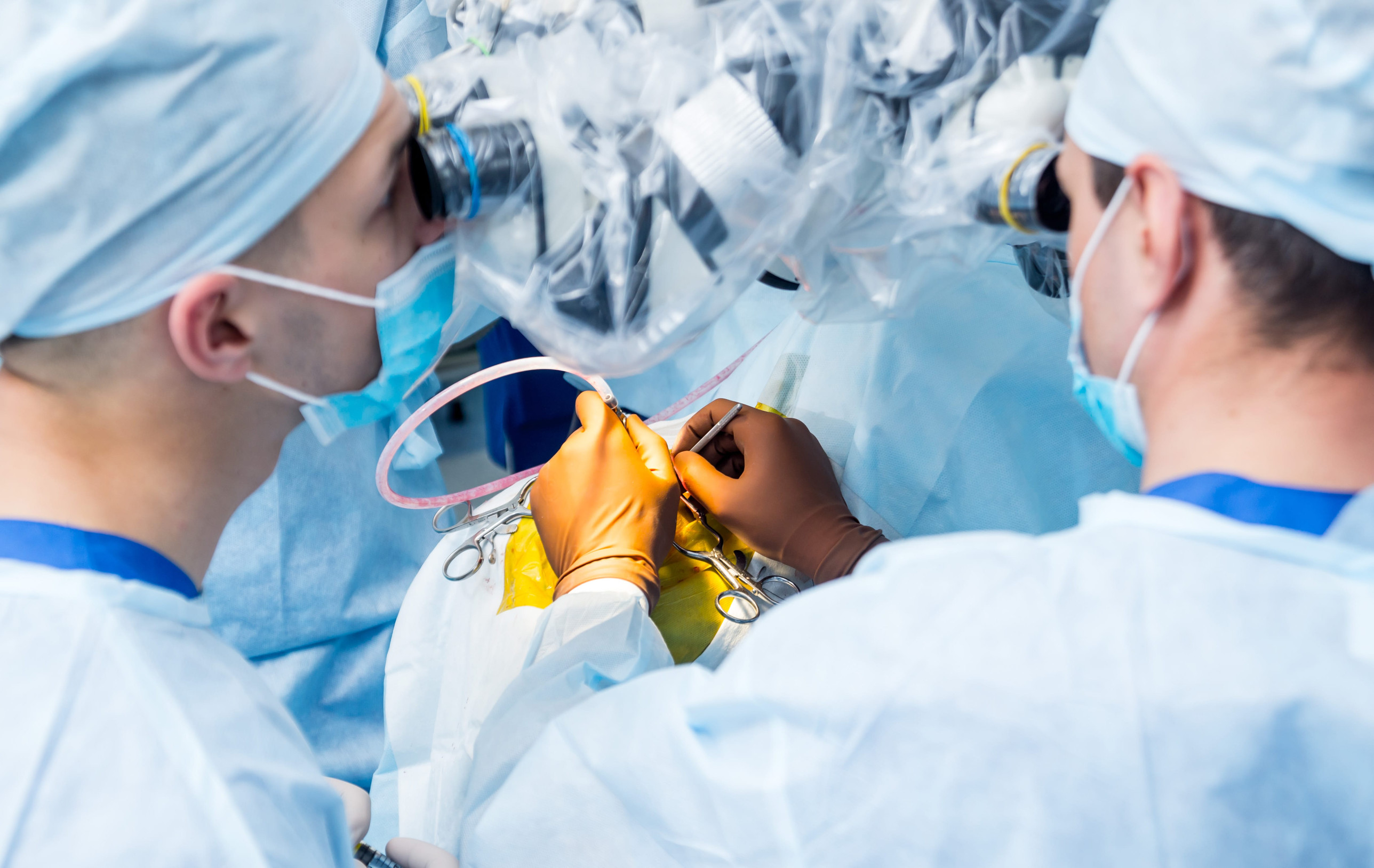
Head and neck microvascular free flap surgery involves transplanting tissue from a donor area of the body such as the arm, back, or leg to a recipient site in the head and neck that has been impacted by cancer, trauma or a congenital birth difference. The donor tissue is transplanted along with its blood supply. An artery provides a source of blood inflow to the tissue and one or more veins allow for drainage of blood from the transplanted tissue. The term "free flap" refers to the tissue transplant that is moved along with its intrinsic arterial and venous blood supply.
The artery and veins that are transplanted with the free flap are connected to recipient arteries and veins in the neck or face to reperfuse the tissue. The vessels are often connected under an operating microscope, which is why the technique is referred to as "microvascular" surgery, or "microvascular free flap" surgery.
Multiple free flap options exist, each with unique characteristics regarding tissue composition, vasculature, and donor site morbidity. Selecting the appropriate flap requires careful consideration of the defect size, location, tissue requirements (skin, muscle, bone, or combinations), and patient factors. Advanced virtual surgical planning for composite defects involving bone helps optimize flap selection and surgical outcomes.
Radial Forearm Free Flap
The radial forearm free flap is one of the most versatile and commonly used flaps for head and neck reconstruction. It provides thin, pliable skin with the potential for sensory reinnervation, making it ideal for intraoral and facial reconstruction. The radial artery provides reliable blood supply. This flap is particularly valuable for floor of mouth defects, tongue and pharyngeal reconstruction, and facial resurfacing. The forearm donor site typically heals well with acceptable functional and aesthetic outcome.
Osteocutaneous Radial Forearm Free Flap
This variation of the radial forearm flap includes bone (radius) along with skin and soft tissue, enabling reconstruction of both soft tissue and bone defects. The included radial bone provides structural support for jaw reconstruction or defects requiring a bone framework. This flap is particularly useful for defects where the thin, pliable skin paddle of the forearm is needed in addition to vascularized bone.
Fibula Free Flap
The fibula free flap is commonly used for mandibular reconstruction due to its long, straight bone segment with excellent blood supply and minimal donor site morbidity. The included skin paddle allows simultaneous soft tissue reconstruction. The fibula's dimensions and shape make it ideal for recreating the mandibular arch, and dental implants can be placed in the fibula bone to restore chewing function. Virtual surgical planning allows precise shaping and positioning to match the patient's anatomy.
Anterolateral Thigh (ALT) Free Flap
The anterolateral thigh free flap provides large amounts of pliable skin and soft tissue with excellent blood supply from the lateral circumflex femoral artery. The ALT flap is highly versatile and can include muscle (vastus lateralis) and/or skin. This flap is ideal for large intraoral defects, pharyngeal reconstruction, and facial resurfacing. The donor site typically heals well with acceptable functional and aesthetic outcome, making ALT a popular choice for many head and neck reconstruction scenarios.
Scapular Angle Free Flap
The scapular angle free flap includes bone from the scapula along with skin and soft tissue when needed, providing excellent bone stock for mandibular or maxillary reconstruction. The scapular bone has favorable dimensions and the angular artery provides reliable vascularization. This flap offers the advantage of a separate skin paddle that can be oriented independently from the bone, allowing optimal positioning for complex three-dimensional defects. The scapular angle is particularly well suited to certain maxillectomy defects. Donor site morbidity is minimal with good functional and cosmetic outcomes.
Latissimus Free Flap
The latissimus dorsi free flap provides large amounts of well-vascularized muscle and skin, making it ideal for extensive soft tissue defects. The thoracodorsal artery provides reliable blood supply to this large, robust flap. Latissimus flaps are particularly valuable for large pharyngeal defects, extensive facial or scalp reconstruction, and situations requiring substantial tissue volume. The donor site heals well with minimal functional impairment.
Iliac Crest Free Flap
The iliac crest free flap includes bone, along with skin and soft tissue. This flap is particularly valuable for mandibular reconstruction, especially in cases requiring substantial bone height and width. The deep circumflex iliac artery provides excellent vascularization. The cancellous bone included with the flap is ideal for dental implant placement. While donor site morbidity is higher than fibula flaps, the iliac crest offers excellent bone dimensions for complex reconstructions.
Medial Sural Artery Perforator Flap
The medial sural artery perforator (MSAP) flap is a thin, pliable flap based on perforating branches of the sural artery in the calf. This flap provides excellent tissue quality for intraoral reconstruction with minimal donor site morbidity. The MSAP flap is particularly valuable for patients with limited alternative flap options or when thin, supple tissue is essential for optimal functional and aesthetic outcomes. The small donor site typically heals with minimal visible scarring.
Free Flap Monitoring
Free flaps are monitored closely in the hospital after surgery. The highest risk period for a blood clot to form in the transplanted vessels is the first 48 hours. The tissue is monitored using a handheld doppler probe in many instances. In some scenarios where the free flap is not easily visible in the oral cavity, face, or neck, an implantable doppler is used to monitor tissue perfusion. Success rates for this sophisticated reconstructive procedure are over 98%.
Expertise and Advanced Planning
Dr. Garg and our surgical team utilize state-of-the-art technology to virtually plan free flap reconstruction, optimizing flap selection, positioning, and shaping for each patient's unique defect. For composite defects involving bone, computer aided design and manufacturing improves surgical precision, reduces operative time, and enhances outcomes. Our multidisciplinary approach includes collaboration with reconstructive, oncologic, and dental specialists to ensure comprehensive care. If you require free flap reconstruction related to cancer removal or trauma, or a congenital facial difference, we invite you to schedule a consultation to discuss your options.